
Can artificial intelligence (AI) be improved by learning how the brain learns?
That question in the mid-2010s prompted the Intelligence Advanced Research Projects Activity (IARPA), a little-known federal agency that’s part of the U.S. intelligence community, to ask whether it would be possible to “reverse-engineer” the brain. This led to the IARPA Machine Intelligence from Cortical Networks (MICrONS) Program, whose goal was to apply the brain’s computational logic to machine learning methods that drive AI.
Now, about 10 years later, the answer to that initial question is closer to “yes.” We got here by building on IARPA’s initial investment, including with support from the National Institutes of Health Brain Research Through Advancing Innovative Neurotechnologies® Initiative, or The NIH BRAIN Initiative®, and other research partners.
On April 10, 2025, the MICrONS Consortium – an international collaboration led by researchers at the Allen Institute for Brain Science, Baylor College of Medicine, and Princeton University – unveiled a fully integrated, highly detailed and dynamic map of a cubic millimeter piece of mouse brain, which enables a vibrant new dialogue between brain science and computational science. The findings were released in 10 research papers in the journal Nature and Nature family journals. Arriving at this moment required the effort of hundreds of researchers, who together achieved technological breakthroughs made possible by the NIH BRAIN Initiative.
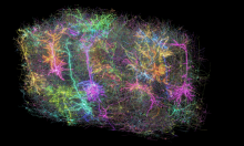
The newly released MICrONS dataset is the largest and most detailed wiring diagram of a mammalian brain ever built. It contains 2.5 miles of axons (the brain’s connection highways) and 523 million connections between 200,000 brain cells in the mouse visual cortex: the brain region that processes information from the visual world. To create the map, before harvesting brain tissue for analysis, the scientists first recorded activity of 76,000 cells in a mouse’s visual cortex while showing the animal video clips of real-life scenes. Then, by overlaying firing activity onto the wiring map, they could deduce what happened, and where, moment by moment.
The results thus show not just which brain cells connect, but also how they fire and communicate electrically in response to many different visual scenes and cues. This work bridges the gap between anatomy and function, a longstanding dream in neuroscience.
Progress has occurred steadily over the past several years. In 2019, BRAIN Initiative-funded scientists began analyzing MICrONS data, including starting a company to facilitate the analysis. This work would later contribute to developing the much larger and ambitious BRAIN CONNECTS program that is currently underway. CONNECTS aims to produce a complete wiring diagram of the entire mouse brain – and of the entire human brain (at a lower resolution).
In 2021, the MICrONS Consortium released MICrONS Explorer: an online, interactive “virtual observatory” of the visual cortex of the mouse brain. This powerful resource allows scientists, developers, and the public to explore the MICrONS dataset. A key contribution to this journey is NIH co-funding and technical guidance for aspects of MICrONS that directly support the BRAIN Initiative’s mission.
Researchers have studied MICrONS data closely using data pipelines, which are automated systems that organize and process large amounts of information using machine learning. They also used advanced tools like high-resolution brain imaging and models of brain circuits. Other BRAIN-funded scientists have used MICrONS tools to reconstruct microscopic images into a three-dimensional map of a tiny chunk of human brain tissue. Published in 2024, this highly detailed anatomical map of healthy brain tissue is a rarity; many studies of human brain anatomy reflect diseased brains. MICrONS offered interactive tools to view and characterize the tiny images, enabled collaborative proofreading by other scientists, and allowed close study of the shapes of neurons and how they connect to each other.
The current BRAIN Initiative CONNECTS research program that grew from MICrONS will take many years to complete, since an entire mouse brain is about 500 times larger than the cubic millimeter of visual cortex analyzed by the MICrONS project. But progress mapping these connections will be an important step toward understanding how our brains make us human. This will be a formidable task: the human brain is roughly 1,000 times larger than a mouse brain, and each of its 86 billion neurons has (on average) more than 1,000 connections. That means the human brain has trillions, if not hundreds of trillions, of connections.
Creating the new MICrONS-enabled brain map also uncovered new and unexpected fundamental knowledge. These insights include wiring diagrams unique to cell types, patterns of how certain brain cells slow down circuits, and a better understanding of how the brain processes information about the visual world. By providing insights into computations underlying how our brains process what we see, the new work will likely also inform AI models through the emerging field of NeuroAI, an area of particular promise for neuroscience. Thus, the work has come full circle, toward improving AI with brain-inspired knowledge.
The highly collaborative MICrONS Consortium, supported initially by IARPA and later by the NIH BRAIN Initiative, is a great example of how open, interdisciplinary interactions within the neuroscience ecosystem can make big things happen. The future of neuroscience, including development of brain treatments that target problem circuits, hinges on the availability of rich datasets like MICrONS that are broadly available to scientists everywhere. In fact, the BRAIN Initiative is supporting efforts to add notes and to proofread the massive dataset, so researchers can focus on the properties and activities of brain cells of particular interest to them.
Many current neuroscience mysteries will likely be addressed by this tiny piece of mouse brain and all the data and insights it has produced and will continue to generate. The best is yet to come, thanks to advances made by MICrONS and other innovative projects supported by the NIH BRAIN Initiative.
With respect and gratitude,
John Ngai, Ph.D.
Director, NIH BRAIN Initiative