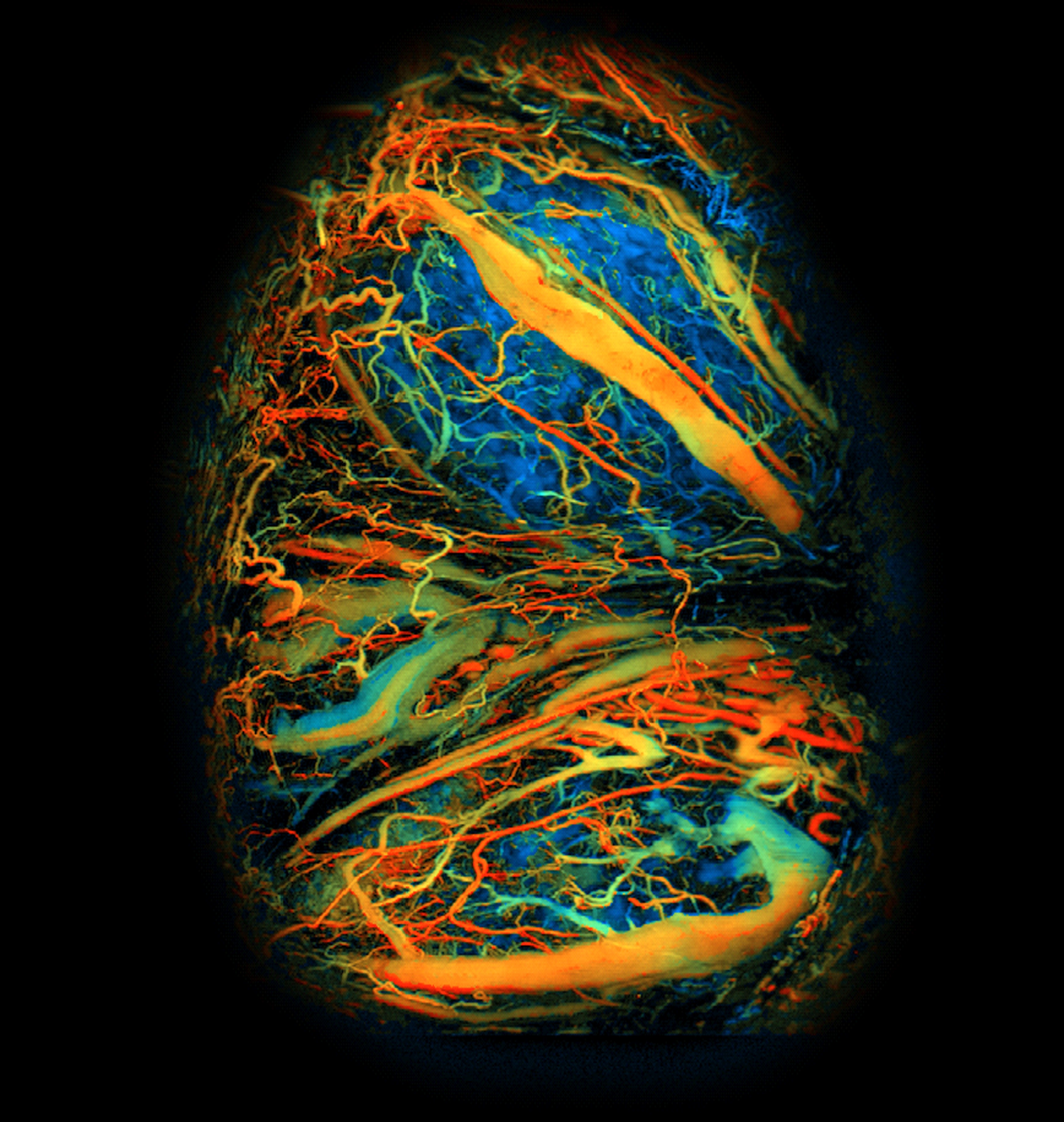
A team of BRAIN researchers have successfully captured real-time, high-resolution images of the developing mouse placenta during pregnancy. These results provide key insights into placental development under both healthy and pathological conditions.
The placenta is a crucial organ in pregnancy, supplying the fetus with oxygen and nutrients essential for survival; however, there is still much about the placenta that we do not fully understand, including the intricacies of its development. Due to its location, ability to move, and the delicate nature of pregnancy, researchers’ ability to obtain detailed images of the placenta in real time have been limited.
A multi-disciplinary group of researchers, funded in part by the BRAIN Initiative, has successfully captured real-time, high-resolution images of the developing mouse placenta during pregnancy. In past studies, the researchers used photoacoustic microscopy (PAM) to record high resolution, high speed, and high throughput measurements of neural activities in freely-behaving animals. In this study, the researchers modified their existing PAM system to make it even faster with a larger field of view. This ultra-fast technique images placenta vasculature and blood oxygenation levels by flashing quick light pulses that generate detectable ultrasound waves after they interact with placental tissues through a window.
Using this state-of-the-art PAM system, the research team monitored mouse placental development in real time in healthy and pathological conditions, such as preeclampsia and alcohol consumption. Preeclampsia is a life-threatening pregnancy complication characterized by a sudden onset of high blood pressure, among other symptoms, that can ultimately lead to maternal and fetal mortality if not properly managed. Researchers injected pregnant mice with lipopolysaccharide (LPS), an inflammatory agent that induces a preeclampsia-like phenotype. Compared with untreated pregnant mice, LPS-treated mice had changes in both oxygenation levels and blood vessel development in the placenta during the early stages of pregnancy. These results suggest preeclampsia pathology may begin at an early stage of pregnancy, warranting further studies.
To evaluate how alcohol consumption can affect the placenta, the researchers administered ethanol into the abdominal cavity of pregnant mice in the second trimester and observed blood flow dynamics. Blood oxygen levels increased within the placenta by almost 30%, lasting for nearly three minutes. These results highlight the sensitivity of the placenta to external stressors and demonstrate how alcohol consumption can have immediate and significant effects on the placental environment.
This work is a notable example of the impact that cross-disciplinary BRAIN-funded technologies can have on organ systems outside of the brain, and could eventually lead to a better understanding of neurological conditions that can result from pregnancy complications. Future studies may include adapting this technique for mammals with longer gestational periods than mice, such as rats and rabbits, and focusing on how environmental factors, such as climate change and water pollution, could impact placental health and outcomes. For more details, please view this science highlight and the full article in Science Advances.
This study was supported by grants from the NIH BRAIN Initiative (RF1NS115581), the National Institute of Biomedical Imaging and Bioengineering (NIBIB; R01EB028143), the National Institute of Neurological Disorders and Stroke (NINDS; R01NS111039), the National Institute of Diabetes and Digestive and Kidney Diseases (NIDDK; R01DK132889 and R01DK119795), the National Institute of General Medical Sciences (NIGMS; R35GM122465), and the National Science Foundation.
Image: Ultra-fast imaging technique captures mouse placenta. This image of a developing mouse placenta was taken during embryonic day 12. Bluer areas have lower levels of blood oxygenation while redder areas have higher levels. Credit: Yao lab/Duke University.
