

NIH BRAIN Initiative grantee Lihong Wang receives award for the development of innovative brain imaging technology.
Lihong Wang, biomedical engineer at Washington University in St. Louis and NIH BRAIN Initiative grantee, was awarded the 2015 Britton Chance Biomedical Optics Award earlier this year by SPIE (the international society for optics and photonics) for his pioneering work towards the development and application of new imaging techniques.
In May, Dr. Wang and his colleagues published a Nature Methods paper describing photoacoustic microscopy—a variation on ultrasound imaging that uses pressure waves to measure biological structures and functions.
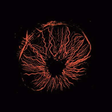
Source: Lihong Wang/Optical Imaging Laboratory/Washington University
Dr. Wang and his group demonstrated the usefulness of this non-invasive technology to perform high-speed, high-resolution imaging of the structure of blood vessels, blood oxygenation levels, blood flow dynamics, and oxygen metabolism of the mouse brain at rest and when electrically stimulated. Photoacoustic microscopy offers several advantages over other imaging techniques commonly used to study the vasculature system of the brain (such as 2-photon microscopy, wide-field optical microscopy, and fMRI), including fast blood oxygenation imaging and fine spatial resolution for defining blood vessel structure. Studying these features of the brain’s vasculature system can provide insight into brain function and the development of diseases such as diabetes and cancer. NPR recently published a story about the potential of this remarkable technique.
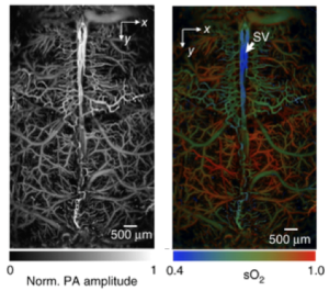
Source: doi:10.1038/nmeth.3336
A technique closely related to photoacoustic microscopy called photoacoustic tomography is the focus of Dr. Wang’s NIH BRAIN Initiative grant in FY 2104. Dr. Wang originally invented photoacoustic tomography (PAT) to detect cancer, but for his BRAIN Initiative grant he has applied the technique to measure the action potentials of large groups of individual neurons. During an action potential, molecules genetically expressed by specific subpopulations of neurons can absorb light, causing them to heat up and release pressure waves that are detected by a device outside the brain and then converted into images of the neuron’s activity. PAT confers several advantages over current techniques for measuring neural activity: it is non-invasive; targets defined subpopulations of neurons; and because brain tissue minimally scatters pressure waves, it can be used for deep penetration to facilitate measurements of whole brain activity.