An official website of the United States government
Official websites use .gov
A
.gov
website belongs to an official government organization in the United States.
Secure .gov websites use HTTPS
A lock
()
or
https://
means you’ve safely connected to the .gov website. Share sensitive information only on official, secure websites.
SHARE:
BRAIN Publication Roundup – August 2019
Image
Noninvasive neuroimaging technology for BCI neuroprosthetics … Genetically-encoded voltage indicator ArcLight maps synaptic activity … Exploring alienation effects of DBS treatment for depression …
Implementing noninvasive neuroimaging technology for BCI neuroprosthetic device control
Brain-computer interfaces (BCI) show promise as therapeutic clinical tools, for example, using signals acquired from intracortical implants to control robotic devices, such as neuroprosthetics. However, invasive recording implants require extensive medical expertise, including high-risk surgical techniques, and are extremely expensive, limiting their use and thus their potential societal impact. While noninvasive counterparts would be ideal in this sense, noninvasive BCI devices have been less effective in capturing and decoding neural signals to the degree needed for functional use. To take on this challenge, Dr. Bin He
. The investigators created two separate training tasks to improve use of a BCI device, asking healthy participants to use motor imagination skills to control a virtual cursor while engaging with a target. One group participated in a traditional BCI discrete trials (DT) task, where they simply had to move a cursor to a static target. The other group participated in a novel, more realistic continuous pursuit (CP) task, using the cursor to chase a continuous, randomly moving target. Analyzing their results, the researchers found that DT training improved performance by 60%. However, the CP task improved performance by 500%, due to its ability to gauge a user’s BCI proficiency and provide more efficient, personalized training. Finally, the researchers tested the practicality of these results by asking participants trained on the CP task to move a robotic arm in 2D instead of a virtual cursor. This task showed a similarly successful performance, indicating a smooth transition from control of a virtual object to a real-world device. Continued improvements in this technology could allow greater access to BCI prosthetics for a wider variety of people with neurological disorders, improving clinical outcomes and restoring once-lost bodily functions without the need for invasive intracortical BCIs.
Using the genetically-encoded voltage indicator ArcLight to map excitatory and inhibitory synaptic activity
Many research experiments currently monitor neuronal activity through calcium imaging, using the concentration of calcium in a cell as an indirect marker for synaptic activity. While this technology has greatly improved scientists’ imaging capabilities, especially through the use of genetically-encoded calcium sensors that are restricted to specific cell types, challenges remain. Calcium imaging provides an indirect and relatively slow measure of synaptic transmission, so it cannot distinguish the nuances of subthreshold or inhibitory changes, ligand-based influx, or spontaneous activity. Dr. Lawrence Cohen
and colleagues at the Yale School of Medicine and the Korea Institute of Science and Technology have been investigating genetically-encoded voltage indicators (GEVIs) as a more direct approach to map excitatory and inhibitory synaptic connections among populations of neurons. The researchers have expanded research on a previously-developed GEVI, called ArcLight. Applying this imaging technology in the CA1 region of the mouse hippocampus in brain slices, they sought to improve resolution and reduce noise in a region that has known excitatory and inhibitory circuits. To achieve this, the investigators created genetic modifications in mice in order to either encode ArcLight in an experimental group, or encode a calcium sensor, GCaMP6f, in a control group for comparison. Fluorescence changes in ArcLight successfully modeled the characteristic pattern of voltage change caused by action potentials, with initial depolarization followed by hyperpolarization, unlike GCaMP6f. This demonstrated that ArcLight could provide a more robust signal, as it was able to represent both excitatory and inhibitory inputs, in ways consistent with the known circuitry of the region. Following this, the researchers confirmed that these changes were due to voltage changes in the cell, rather than spontaneous synaptic activity. Contrastingly, the activity of GCaMP6f could not represent inhibitory inputs or hyperpolarization and had a much slower flux and response time. Collectively, these results demonstrate a viable approach to mapping neural circuitry, combining structural mapping, which alone does not confirm functional connections, with functional imaging with GEVIs. Future advances in this technology bring hope of creating a full connectome map of the brain, which could improve our understanding of how circuits function, and how their dysfunction impacts human health.
Image
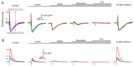
Comparison of activity between ArcLight- and GCaMP6f-expressing neurons. (Top) Representation of fluorescence changes in ArcLight-expressing neurons in response to electric stimulation. ArcLight-expressing neurons show a quick depolarization and hyperpolarization, characteristic of an action potential. These effects are blocked using specific receptor blockers and neurotoxins, demonstrating that the activity of ArcLight responds specifically to voltage change, not spontaneous activity. (Bottom) Representation of fluorescence changes in GCaMP6f-expressing neurons in response to electric stimulation. GCaMP6f-expressing neurons show a slower depolarization without hyperpolarization, demonstrating an inability to represent inhibitory inputs. This is further demonstrated by control experiments, which show no unique responses to blockers for inhibitory signal receptors.
Exploring the possible alienating effects of DBS treatment for depression
Aligning with the BRAIN Initiative’s vision of advancing novel neurotechnologies and their role in medicine, Deep Brain Stimulation (DBS) has proven effective for certain neurological disorders, and has shown some promise as a treatment for several other neurological and psychiatric conditions. While DBS has shown potential in treating symptoms of depression, previous research has identified possible ethical concerns. For example, some studies have reported patients feeling disconnected from many aspects of their life when considering their states before and after treatment, experiencing alienation and estrangement. As a result, Dr. Gabriel Lazaro-Munoz
. In their analysis, the researchers made the distinction between alienation from self and alienation from other objects, such as one’s work and personal relationships. They found that many patients were unable to identify with themselves or felt disconnected from targeted aspects of their identity or agency, as a form of self-alienation after DBS treatment for depression. These patients claimed that they “don’t recognize” themselves, felt “like a machine,” or believed their “body is cured” but their “mind is still sick” after DBS treatment, experiencing mental states not characteristic of themselves. Beyond this, some patients failed to identify with previously enjoyed work or relationships, with varying degrees of withdrawal. Finally, the researchers identify patients with depression as a rich source for future evaluations of the relationship between DBS treatment and alienation. Since many symptoms of depression, such as anhedonia, dysphoria, and social withdrawal, can be described as varying degrees of alienation from different objects, patients who have undergone DBS can understand the differences between their feelings of alienation before and after treatment, allowing researchers to distinguish between the specific effects of DBS and general symptoms of depression. Dr. Lazaro-Munoz and his colleagues call for continued research, since alienation can deprive an individual of important improvements in their quality of life, despite potential successful clinical outcomes. While the increased utilization and effectiveness of neurotechnologies like DBS in medicine hold exciting therapeutic potential for millions of people suffering from brain-based disorders, this group has highlighted important neuroethical considerations that can inform future medical research, policy, and practice.