An official website of the United States government
Official websites use .gov
A
.gov
website belongs to an official government organization in the United States.
Secure .gov websites use HTTPS
A lock
()
or
https://
means you’ve safely connected to the .gov website. Share sensitive information only on official, secure websites.
SHARE:
BRAIN Publication Roundup – July 2017
Method to overcome light scatter in optical imaging distinguishes moving objects with high fidelity… Novel analysis method improves accuracy of neural network models… Improved imaging technique reveals synaptic transmission at quantal resolution in fruit fly larvae
Phase retrieval methods in optical imaging allow for successful imaging of moving targets through scattered media
A major challenge of optical imaging is the scattering of light, which leads to lower resolution, poorer image quality, and shallower depths of images, especially those involving biological tissue. Researchers have tried to overcome these effects by filtering out repeated scattering of light to measure only the un-scattered or minimally scattered photons. However, such methods fail to address limitations in depth of imaging because they detect only the minimally scattered photons, which are difficult to observe at greater distances. Optical imaging approaches that incorporate information from the scattered photons often require long acquisition times, a reference source (also known as a “guide star”), and dark-field conditions. Recently, Dr. Changhuei Yang and his group
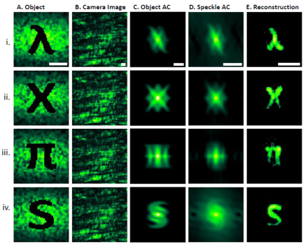
at California Institute of Technology have developed a successful method to image moving objects through scattering media. Using temporal and angular correlational analyses from the scattering process, they distinguished objects from their backgrounds, permitting imaging of moving targets that would otherwise have been hidden due to the scattering media. This technique can be used in both dark-field and light-field scenarios, allowing the imaging of non-emitting, transmissive, and reflective non-static samples. This exciting new method could be extended to biological tissue, such as imaging deep brain structures in freely moving animals, and possibly other applications, such as moving objects in dense environments like fog or underwater.
Image
A) Objects were hidden behind a scattering medium and moved 1.5 mm between each acquisition (4 acquisitions per image). B) The raw camera images were blurry and indistinguishable due to the scattering of light from the background. Images remain indistinct based on C) object autocorrelations and D) speckle autocorrelation analyses. E) Using a phase retrieval algorithm, each object was successfully reconstructed.
Functional imaging analysis incorporates connectivity and structural relatedness to improve modeling of brain functional networks
Functional network modeling from resting-state functional magnetic resonance imaging (fMRI) helps clarify how the brain functions normally and could identify biomarkers of neurological and psychiatric disorders. Often, models represent regions of interest (ROIs) in the brain as “nodes” with the functional connectivity between these regions delineated “edges.” At the University of North Carolina, Chapel Hill, Dr. Dinggang Shen’s group
developed a new method, connectivity strength-weighted sparse group representation, to model brain functional networks. Like other methods, the team relied on pairwise correlations between ROIs to infer connectivity and made the neurologically reasonable assumption of sparse networks. From BOLD signals, the model assumed that brain regions have ‘first order/direct” interactions with only a few other regions and reduced spurious connections. However, unlike previous methods, the model made use of information on variations in connectivity strength and groupings of similar ROIs. Thus, the team combined pairwise correlations to measure interactions across multiple ROIs, weighted functional connectivity strength to account for network sparsity, and incorporated ROI group structures (a set of regions with similar characteristics) into one model. Using fMRI data from individuals with mild cognitive impairment (MCI), an early indicator of Alzheimer’s disease or another dementia, and from normal controls, the group tested the model on seven performance metrics, including accuracy, sensitivity, and specificity. The team found that their model successfully classified the data as MCI or control better than other brain network connection models with almost 85% accuracy compared to 65%. These results provide validation that this model can identify biomarkers of MCI, which could guide early interventions for Alzheimer’s disease. Shen’s group intends to improve the grouping of related structures in the model further and apply to different brain disorders and diseases.
Image
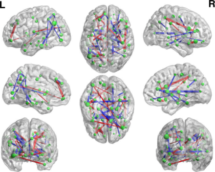
This figure shows the pattern of nodes and edges that are most likely to indicate MCI classification, an early marker of Alzheimer’s disease. The nodes, represented by green spheres, mark regions of interest and the edges, represented by red and blue lines, identify the connectivity between the regions. These nodes and edges were consistently selected by the new method of connectivity strength-weighted sparse group representation. Note that increased line thickness is represented by increased connectivity as measured by fMRI BOLD response.
Novel quantal resolution imaging technique advances understanding of input-specific plasticity and homeostasis at the Drosophila larval neuromuscular junction
Synapses, including those using glutamate as a neurotransmitter, vary greatly in pre- and post-synaptic transmission properties. How differences in pre-synaptic release characteristics, short term plasticity, and homeostatic stability-promoting regulation relate to one another and to this diversity is poorly understood. At the University of California, Berkeley, Dr. Ehud Isacoff
and his group used the Drosophila larval neuromuscular junction (NMJ), a model system for studying glutamatergic transmission, to better understand these mechanisms. They used novel quantal resolution imaging to study the role of input and synapse specificity in the regulation of basal synaptic strength, plasticity, and homeostasis. This technique relies on transgenic Drosophila that express a genetically-encoded calcium indicator, SynapGCaMP6f, allowing for the imaging and quantification of post-synaptic transmission without voltage clamping. Glutamate is released during basal and evoked synaptic events following increases in calcium, revealed by SynapGCaMP6f as changes in fluorescence. Thus, the researchers used fluorescent imaging to examine the probability, frequency, and amplitude of quantal spontaneous, and activity-dependent synaptic transmission of two different synapes onto muscle, those from Ib and Is neurons. By applying this technique with and without motor input, the researchers discovered that Ib and Is synapses have different basal synaptic release characteristics and activity-dependent synaptic modulation at the NMJ. Strikingly, homeostatic compensation in synaptic strength occurred only in the 1b synapses. BRAIN-supported advances in imaging techniques such as this continue to give unprecedented insight into how the nervous system carries out computations and can be expanded across model systems.
Image
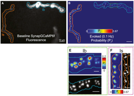
Panel A. Baseline fluorescence of the calcium indicator in Ib (blue) relative to Is (orange) at the NMJ. Panel B. Evoked synaptic activity at Ib (blue) relative to Is (orange) at the NMJ. Warmer colors indicate an increased probability of glutamate release. Panels E and F. High-magnification comparison of quantal evoked activity for each neuron.