An official website of the United States government
Official websites use .gov
A
.gov
website belongs to an official government organization in the United States.
Secure .gov websites use HTTPS
A lock
()
or
https://
means you’ve safely connected to the .gov website. Share sensitive information only on official, secure websites.
SHARE:
BRAIN Publication Roundup – June 2017
Image
BRAIN Initiative team pushes the limits of functional magnetic resonance imaging (fMRI) for human brain… A novel tool to manipulate gene function in specific cell types… Understanding the functional diversity of retinal bipolar cells…
Advancements to functional imaging technique result in ultra-high resolution capture of human cortical columns
Despite numerous advances in fMRI technology, most components are optimized for the entire body. This makes safe, ultra-high resolution (UHR) imaging of columnar organization throughout the cortex of the human brain nearly impossible. At the University of California, Berkeley, Dr. David Feinberg
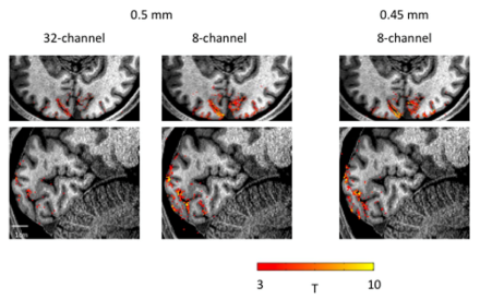
and colleagues applied updates to magnetic gradients, receiver arrays, and pulse sequences of simultaneous multi-slice echo planar imaging fMRI to achieve UHR imaging of human ocular dominance columns. Focusing particularly on a prototype receiver array (8 channels with 4 cm diameter coils), the group systematically describes the changes necessary to achieve the higher signal-to-noise ratio required to attain ~0.5 mm imaging resolution in 3 dimensions. Finally, the researchers display their updated UHR system compared to commercially available technology when mapping ocular dominance columns in three subjects shown visual stimuli in the scanner. The group notes their findings are part of a growing set of 3D imaging studies, moving to leverage fMRI to understand neural circuitry by revealing activity of distinct cell populations in different cortical layers. They postulate that this 3D imaging technique could eventually progress from 0.5 mm to 300-400 µm resolution fMRI of the entire human brain.
Image
Brief fMRI scans from a visual activation paradigm in a human subject reveal enhanced cortical activation at 0.5 mm spatial resolution in the prototype receiver array (8-channel with 4 cm diameter loops) compared to commercially available technology (32-channel). The prototype array accurately measured activation at 0.45 mm resolution as well, further illustrating improved signal-to-noise ratio.
New technique developed for controlling gene function in distinct cell types in the fruit fly
The ability to manipulate genes in specific cell types is critical for understanding circuit function and dysfunction. Unfortunately, there are limitations for the currently available tools, including off-target effects (i.e., modifying genes other than the targeted gene), incomplete inactivation of the targeted gene, requiring cell division (which restricts the time during development when the gene can be targeted), and incompatibility with model systems like the fruit fly Drosophila melanogaster, which is a principle model for studying neural development and function. At Stanford University, Dr. Thomas Clandinin
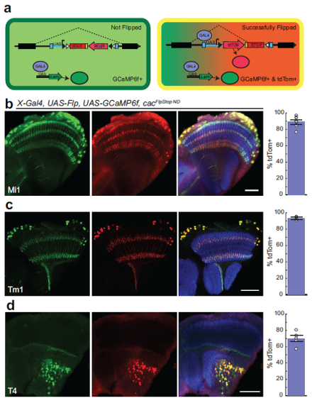
and colleagues have developed a tool called FlpStop, which can completely disrupt targeted genes, as well as rescue gene expression of the disrupted genes, in differentiated and undifferentiated cells in Drosophila. FlpStop uses endogenous mechanisms within cells and a process called insertional mutagenesis to completely inactivate/disrupt the normal function of a gene. The FlpStop insertion is tagged with a fluorescent protein so that mutant cells can be visualized, and it can be inserted in both non-disrupting and disrupting orientations. The team successfully disrupted gene function in six out of eight of the genes that were tested and confirmed that the non-disrupting orientation did not interfere with gene function in any case. They successfully labeled the FlpStop inserted genes in three different cell types in the Drosophila visual system (Mi1, Tm1, and T4). Finally, the group effectively combined FlpStop with in vivo calcium imaging. The findings suggest that FlpStop represents a promising and powerful new tool for investigating gene function in specific cell types.
Image
(a) Schematic of the experimental design for testing the effectiveness of the FlpStop insertion. Drosophila Gal4 driver lines were used, whereby three distinct cell types in the visual system were targeted: (b) Mi1, (c) Tm1, and (d) T4. The full expression of each Gal4 driver line is labeled green while the combination of Gal4 (in green) and the successfully-expressed FlpStop gene (in red) together appear yellow, demonstrating 70%-93% overlap depending on the cell type.Studying the functional diversity of bipolar cells improves understanding of neuronal processing in the visual system
One core goal of the BRAIN Initiative is to better understand brain circuitry and the role of different cell types in these brain circuits, in both healthy and diseased brains. A relevant area of focus is retinal neurons and their ability to encode visual stimuli for the brain. While the anatomy and genetics of retinal bipolar cells are well characterized, their functional diversity is incompletely understood. In an article in Nature, BRAIN awardee Dr. Thomas Euler
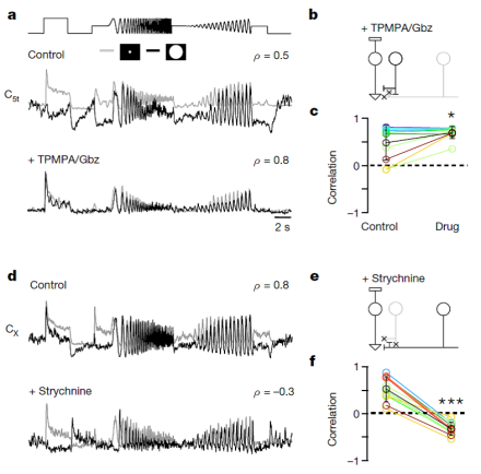
and colleagues used two-photon imaging to examine the effects of amacrine cell activity (a type of retinal interneuron) on the output of bipolar cells in the mouse retina. Through exposing different areas of the retina to light, the researchers found that functionally opposite signals, such as those used to describe ON and OFF bipolar cells, exist at the same layer of the retina, suggesting that the retina structure is more complicated than previously thought. Furthermore, inhibition of amacrine cell activity led to increases in the functional diversity of bipolar cells. The team determined that a bipolar cell’s output is determined by a combination of excitatory input to the dendrite and amacrine cell input to the axon, which ultimately allows for temporal encoding in the visual system. These important findings give us a better understanding of the visual system and of the mechanisms through which different neuronal cell types communicate with one another.
Image
Local (gray) and full-field (black) output responses of bipolar cells in both control and drug conditions. TPMPA/Gbz blocks GABAergic amacrine cell activity, while strychnine blocks glycinergic amacrine cell activity. Blocking amacrine cell activity led to opposite responses from bipolar cells compared to control conditions.