An official website of the United States government
Official websites use .gov
A
.gov
website belongs to an official government organization in the United States.
Secure .gov websites use HTTPS
A lock
()
or
https://
means you’ve safely connected to the .gov website. Share sensitive information only on official, secure websites.
SHARE:
BRAIN Publication Roundup – September 2019
Image
Manipulating neural activity while measuring physiology in awake mice … Discovery of the brightest green fluorescent protein homolog to date … Assessing mouse neural dynamics with optoacoustic imaging … Establishing new open cloud services for brain data …
Repeated imaging of awake mice with multiple imaging modalities yields neural, microscopic, and mesoscopic data
Functional magnetic resonance imaging (fMRI) is widely used to non-invasively study human brain activity. However, how the underlying physiology of individual brain and blood vessel cells generates the fMRI signals is not well understood. Performing invasive cellular measurements that are not currently possible in humans and non-invasive imaging in mice provides a bridge to understand the cellular underpinnings of fMRI. Researchers have now demonstrated the feasibility of doing repeated non-invasive and invasive imaging in unanesthetized mice. Dr. Anna Devor
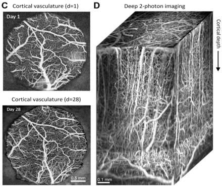
and colleagues at the University of California, San Diego devised a protocol to image awake mice using various imaging techniques that ultimately allows for multiscale data generation. Each mouse was imaged using two-photon microscopy, laser speckle contrast imaging, and blood-oxygen level-dependent (BOLD) fMRI. There were several goals of this study, including ensuring that the cranial window implant remained clear, preventing signal artifacts, maintaining the stability of the headpost, allowing for repeated imaging over weeks and months, and conducting behavioral experiments. Genetically-engineered mice expressing inhibitory neurons with the channelrhodopsin-2 optogenetic actuator protein received several different stimuli to compare neural activity across imaging modalities. Using both one and two-photon imaging, insights on the activity of neurons, glial cells, vascular cells, and overall brain metabolics were determined at submicron resolution, as well as glucose and oxygen consumption. BOLD fMRI imaging was also sensitive enough to detect stimulus conditions in specific cortical regions. The authors found a notable increase in neural activity in fully awake versus sedated mice, likely due to decreased blood flow of mice under anesthesia. These results showcase a feasible imaging protocol in which multiple levels of brain data can be elucidated from awake mice, and this protocol could be extended to complex behaviors, such as sensory discrimination or attention. Dr. Devor and her team’s results exemplify how different brain imaging techniques can be synthesized to create a more dynamic and fundamental understanding of the brain.
Image
Two-photon imaging of awake mice through long-term glass cranial windows. (C) Imaging of cortical vasculature on Day 1 and Day 28 after cranial window implantation. (D) An image stack generated from two-photon imaging of cortical depth via cranial window.
Unraveling a “jellyfish’s secret” leads to the characterization of nine previously unknown fluorescent protein homologs
Green fluorescent protein (GFP) and its variants have revolutionized science because of their ability to showcase living complex biological systems at high levels of resolution – including the mammalian brain. However, fluorescent proteins used for research have only been derived from a small number of marine organisms, such as corals and jellyfish. Most fluorescent proteins used in scientific applications arise from the sea anemone Entacmaea quadricolor or the coral Discosoma, because of their ability to emit at longer wavelengths of light. Dr. Nathan Shaner
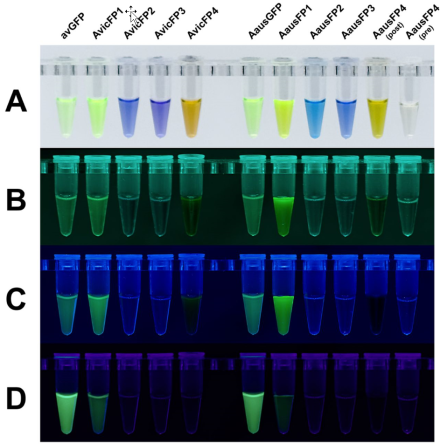
. The overall goal of this study was to overcome the limitations of the known fluorescent protein scaffolds by identifying, characterizing, and bioengineering fluorescent proteins from more diverse marine organisms with little homology to commonly used fluorescent proteins. After analyzing the transcriptome of A. cf. australis (an Aequorea species most similar to Aequorea australis) in the Great Barrier Reef, the authors identified the brightest fluorescent protein to date – named AausFP1 – with a more than five-fold molecular brightness than enhanced GFP (EGFP). Returning to San Diego, the authors discovered that Aequorea victoria, on exhibit at a local aquarium, also expressed orthologs of AausFP1 that they originally identified in A. cf. australis. Beyond green emitters, these investigations discovered a wide variety of other potentially useful fluorescent proteins, including, for example, purple and blue pigmented chromophores with absorbances ranging from green to the far red. Importantly, several fluorescent variants were superior scaffolds than GFP, meaning that they are more stable in various physiological mammalian conditions (e.g., 37°C). Dr. Shaner and his colleagues’ work emphasizes the importance of characterizing unknown species with the capacity to advance science that may not exist in the future. These discoveries also highlight the value of a highly interdisciplinary approach, including field collection, molecular biology, next-gen sequencing, bioinformatics, protein engineering, microscopy, x-ray chrystallography, and phylogenetics.
Image
Fluorescent proteins purified from different Aequorea species. Different fluorescent proteins under (A) white light, (B) 505 nm, (C) 480 nm, or (D) 400 LED illumination. Note that AausFP1 shares 53% homology to avGFP and is currently the brightest fluorescent protein characterized.
Real-time volumetric measurements using optoacoustic imaging of the mouse brain produces macroscopic snapshots of neural activity
Although indirect methods can image brain-wide activity in the mammalian brain, direct methods for visualization of real time activity in vivo have been challenging. Optical methods, for example, may require invasive methods for deeper structures and are limited in area of coverage. Dr. Daniel Razansky
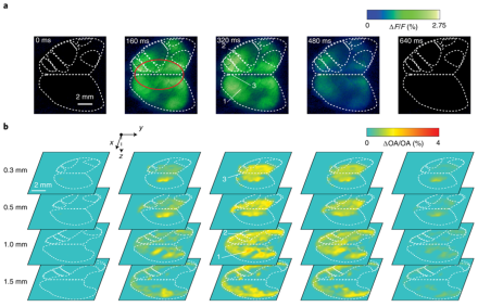
and colleagues at the University of Zurich in Switzerland have developed methods to image whole-brain neural activity in real-time using hybrid optoacoustic imaging. Optoacoustic imaging involves using ultrasound waves that are generated from transiently absorbed light. This allows for deeper imaging of the tissue of interest with fields-of-view near 2 cm3 and spatial resolutions of 150 µm – effectively covering the entire mouse brain. The overall goal of this work was to determine the feasibility of real-time volumetric brain imaging of mice with genetically-encoded calcium indicators. These mice express either fast or slow variant calcium indicators that detect neural activity down to the single neuron level. The authors successfully imaged global calcium activity in the brain as well as hemodynamic responses. Of importance, optoacoustic imaging allowed for the capture of brain processes at different time scales and at depths not previously achievable with other noninvasive imaging techniques. Despite formidable background from hemoglobin absorption in brain tissues, optoacoustic imaging was nevertheless sensitive enough to distinguish both slow and fast changes in neuronal calcium signaling. Additionally, optoacoustic imaging recorded stronger calcium changes than the typical fluorescence activity associated with the calcium indicators. Dr. Razansky and his team’s results showcase the practical utility of optoacoustic imaging for viewing deeper brain regions and for conserving spatiotemporal information on vastly different scales, paving the way for even deeper imaging of the mammalian brain in potentially clinically relevant settings.
Image
Optoacoustic imaging of neural activity in GCaMP6f-expressing mice in response to a hind paw stimulus. Sequence ranging from t=0 ms of a hind paw stimulus to t=160 ms, t=320 ms, t=480 ms and t=640 ms of (A) fluorescence recorded brain activation maps and (B) 4D optoacoustic recorded brain activation maps at different depths ranging from 0.3 mm to 1.5 mm. Dashed white lines represent functional brain regions.
Establishment of an open diffusion data derivative (O3D) repository of brain data encourages integration, data upcycling, and reproducibility among researchers
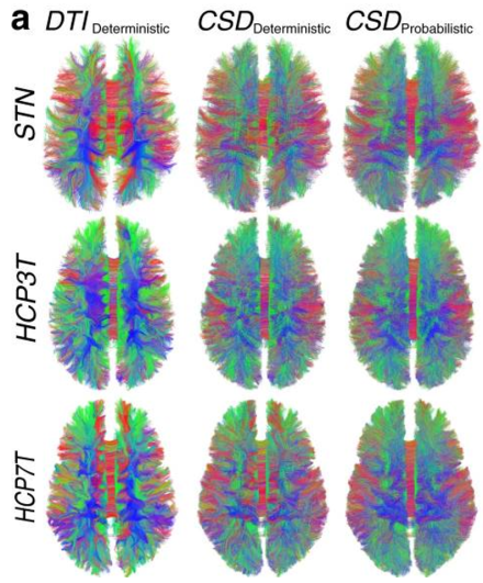
Large scale neuroimaging projects, including the Human Connectome Project, the Alzheimer’s Disease Neuroimaging Initiative (ADNI), the Adolescent Brain Cognitive Development (ABCD) Study and many others have provided resources for data sharing, reuse, standardization, and secondary data analyses. Dr. Eleftherios Garyfallidis
, was created using diffusion-weighted magnetic resonance imaging (dMRI) data and tractography allowing for structural, anatomical, macroscopic, and microscopic details of the human brain to be preserved under one single digital object identifier (DOI). The O3D repository houses a collection of dMRI files from 12 brains; 3 separate datasets; over 300 tractograms; over 7,000 segmented major tracts, and over 700 matrices (connectivity networks). Thus, researchers who use the O3D repository can choose to upload new data using the same data analysis pipelines. Alternatively, individuals can analyze new data by downloading separate data derivatives in a stepwise fashion. The O3D repository differs from similar data projects as it allows for brain data to be tested and retested for reproducibility purposes. The work of Dr. Eleftherios Garyfallidis and colleagues facilitates collaboration between trainees at all career levels and among members of different scientific disciplines. This work, supported by a joint initiative of the National Institutes of Health and National Science Foundation known as the Collaborative Research in Computational Neuroscience (CRCNS) program, and with support from the National Science Foundation and Microsoft, propels the field of neuroscience into the future of making data understandable, interactive, and accessible for all. For more information, please see an Indiana University
Tractograms of the whole human brain. Visualization of whole-brain tractograms generated from three datasets: Stanford University (STN) and the Human Connectome Project (HCP3T and HCP7T). Each dataset uses various reconstruction models, including the deterministic model (DTI), constrained-spherical deconvolution (CSD) deterministic model, and CSD probabilistic model.