

Research in Drosophila reveals context-dependent visual responses in a brain region thought to be involved in navigation during flight
Traditionally, due to technical limitations, most in vivo neuroscience research has taken place in sedated or otherwise restrained animals. While data obtained in such experiments is highly informative, this approach is problematic for studies that investigate the neural basis of behavior. Fortunately, recent technical advances in the miniaturization and sensitivity of electrophysiology and optical imaging instruments have given researchers the ability to monitor the activity of individual neurons, as well as entire populations of neurons, during locomotive behaviors such as walking, running, and flying. New techniques such as these, and the scientific discoveries they enable, are a central focus of the BRAIN Initiative.
BRAIN Initiative awardee Michael Dickinson, PhD, and his colleague Peter Weir, PhD, both of the California Institute of Technology, described in a recent PNAS paper a virtual reality setup in which fruit flies (Drosopohila) were tethered, yet allowed to flap their wings and fly in place. A variety of visual stimuli—including thick stripes moving from left to right or small dots receding into the distance or rushing toward the animal—were presented to the flies to give them a sense of motion as well as visual cues important for guidance and stability during flight.
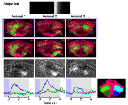
During periods of both rest and flight, the researchers used multiphoton imaging of genetically encoded calcium indicators to record activity of neurons within three discrete regions in a brain area called CX—an area known to be involved in both sensory processing and motor control. All three regions of the CX contained neurons that responded to the visual stimuli. However, only one region, called the fan body, responded selectively during flight and not during rest. This result suggests that the fan body is important for visual control of flight. Additional analyses suggested that two subregions of the fan body play a specific role in controlling heading in the horizontal plane during flight.
Finally, the researchers performed a screen of thousands of transgenic Drosophila mutants to identify eight functional classes of fan body neurons that became activated during each of the different visual pattern types presented by the researchers.
The fact that the functional connectivity of the neural circuits under study depended on the animals’ behavioral state points to limits in what can be learned solely through anatomical studies of neural connections.