Neural circuitry of brain regions involved in decision-making

Image
NIH logo

BRAIN Initiative grantee Dr. Carlos Brody co-authored a Nature paper that explores how distinct neural circuits contribute to the brain’s process of making decisions based on weighing evidence.

Dr. Carlos Brody, an HHMI investigator at Princeton University, and colleagues have published a paper in Nature that pushes the envelope of studying how the brain works to make informed decisions. This research supports the aim of Dr. Brody’s BRAIN grant, which focuses on studying the neural circuit dynamics that underlie working memory – specifically, the accumulation of evidence over time scales of seconds, a type of working memory critical for decision-makingThe work also addresses the broad goal of The Brain InitiativeSM: to understand the circuits and patterns of neural activity that give rise to mental experience and behavior, by beginning to unravel the immense complexity of neural circuits and dynamic neural coding.

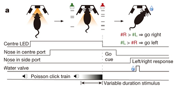
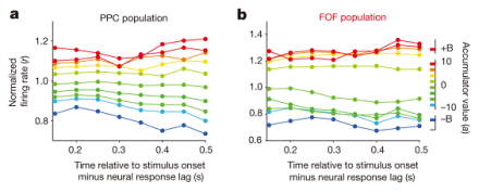
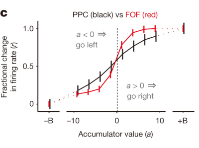
The authors trained laboratory rats to listen for accumulating auditory signals, and then make a decision to turn to the side (right or left) that had played the greatest number of clicks for a reward. By recording neuronal firing rates, first author Hanks et al. show that the posterior parietal cortex (PPC) builds the weight of evidence for which is the correct side to choose, whereas an area of the prefrontal cortex (frontal orienting fields, FOF) provides the ‘go’ signal for the decision. In support of this observation, the authors utilized optogenetics to show that FOF inhibition impacts the decision-making behavior only when applied around the time of the decision, not during early accumulation of auditory evidence. This is an elegant use of a simple experimental system to explore how the brain makes complex decisions.

Image
Subjects hear simultaneously presented trains of randomly timed clicks, one train coming from a speaker to their left, the other from a speaker to their right. At the end of the trains, the subjects have to decide which side played the greater total number of clicks.
Image
These graphs show the relationship between PPC or FOF neural activity and accumulated auditory evidence, aligned to the stimulus onset minus the neural response lag. Colors represent the weight of accumulated evidence, and each vertical slice corresponds to a tuning curve for n = 93 neurons (a) or n = 128 neurons (b). For the PPC, the map of evidence accumulation leading to a response is graded and relatively stable over time.
These graphs show the relationship between PPC or FOF neural activity and accumulated auditory evidence, aligned to the stimulus onset minus the neural response lag. Colors represent the weight of accumulated evidence, and each vertical slice corresponds to a tuning curve for n = 93 neurons (a) or n = 128 neurons (b). For the PPC, the map of evidence accumulation leading to a response is graded and relatively stable over time. For the FOF, the map is relatively stable over time, but the responses cluster based on whether the weight of evidence is positive or negative.


Image
This graph shows average (± s.e.m.) fractional change in firing rate as a function of the accumulated weight of evidence. PPC shows a smoothly graded function, while FOF shows a steeper sigmoid with responses clustered around positive or negative weight of evidence.
This graph shows average (± s.e.m.) fractional change in firing rate as a function of the accumulated weight of evidence. PPC shows a smoothly graded function, while FOF shows a steeper sigmoid with responses clustered around positive or negative weight of evidence.

Latest from The BRAIN Blog

The BRAIN Blog covers updates and announcements on BRAIN Initiative research, events, and news. 

Hear from BRAIN Initiative trainees, learn about new scientific advancements, and find out about recent funding opportunities by visiting The BRAIN Blog.

This site is protected by reCAPTCHA and the Google Privacy Policyand Terms of Serviceapply.
Image
black and white image of people working on laptops at a counter height table on stools at the annual BRAIN meeting