
As we prepare to host the 2026 BRAIN Initiative Conference, we are excited to launch this year's photo and video contest. This unique contest is an opportunity to showcase the stunning, colorful, and inspiring images captured by today's advances in neurotechnology. We're excited to check out your creative fusions of art and science!
Learn More and Submit Your Photos and Videos! Submissions are due by Thursday, July 2, 2026 at 11:59 PM Eastern Time.
Past Winners
Explore the winners of previous year's entries.
2025
2024
BRAIN at 10 Video Finalist
Photo Winners
First place
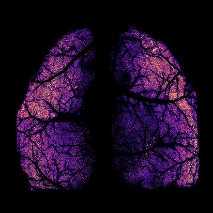
Whole-cortex scale in vivo two-photon imaging with single-cell resolution in mice
Using the newly invented Light Pipe Microscope array, the whole-cortex scale in vivo two-photon imaging is shown with single-cell resolution in CX3CR1-eGFP mice.
By Zongyue Cheng, Jianian Lin, and Meng Cui, Purdue University
Second place
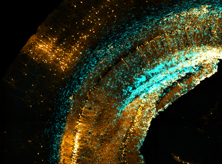
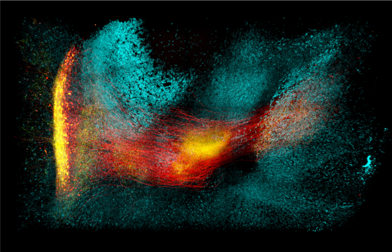
Chaos circuitry
Rabies virus-mediated transsynaptic tracing reveals a vast landscape of neurons (orange) in the mouse brain that synapse with diffusely infiltrative glioblastoma cells (blue), a lethal brain cancer.
By Yusha Sun and Xin Wang, University of Pennsylvania
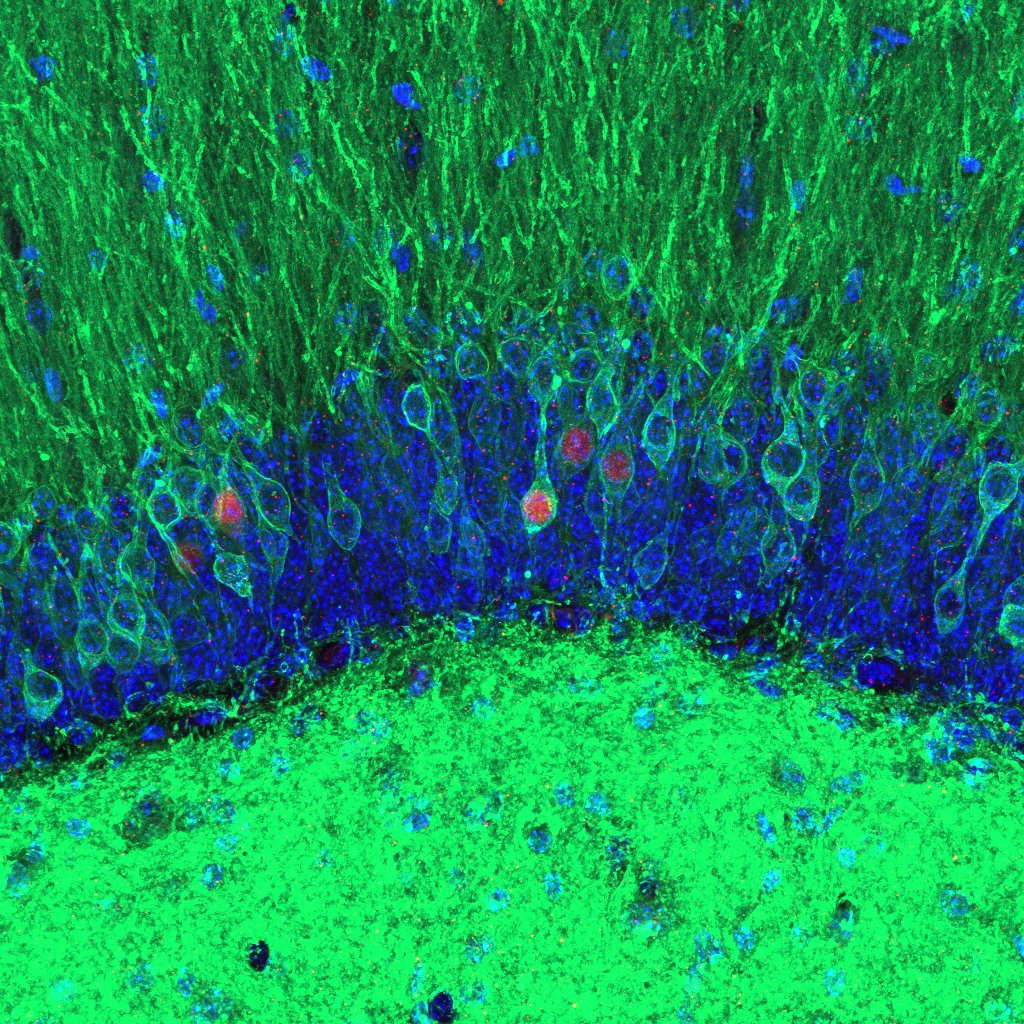
Third place
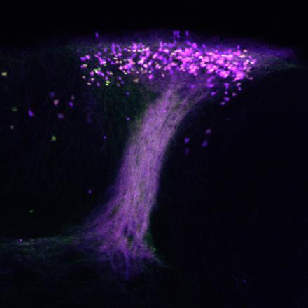
Willow on the Island of Calleja
Exploring the formation of long-term olfactory memory in the mouse brain, stored amongst the neurons of the Island of Calleja (green-violet).
By Lee O. Vaasjo and Maria J. Galazo, Tulane University
BRAIN at 10 Photo Finalist
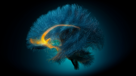
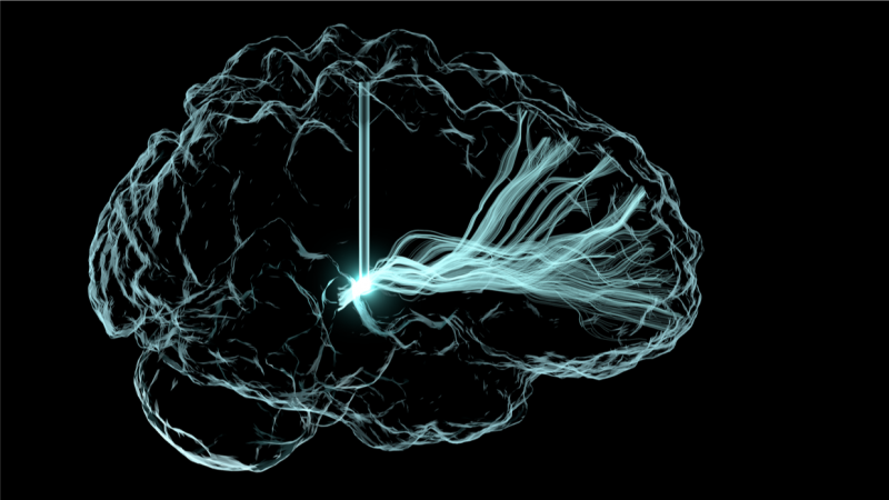
Decoding Depression
Diffusion tensor imaging of the 3D white matter structure in a patient living with depression and using deep brain stimulation. This image highlights the target network for stimulation and decoding the depression state from chronic recordings.
By Chris Rozell, Mike Halerz , Ki Sueng Choi, Helen Mayberg, and Mike Halerz, Georgia Institute of Technology, Icahn School of Medicine at Mt. Sinai and TeraPixel, Inc.
2023
Video Winners
Photo Winners
First Place
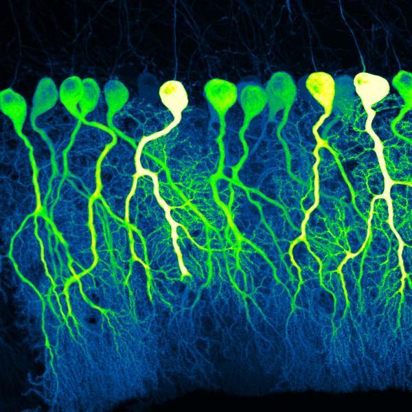
Dark Commute at 4am
In the darkness of a confocal microscope room, bright fluorescent dyes reveal Purkinje cells winding their way through the tissue of the cerebellum. These complex, branching cells play roles in learning and memory. The cells in this photo, taken from sections of mouse cerebellum, resemble pre-dawn commuters on the highways of the brain as they travel towards their eventual targets.
By Silas Busch, University of Chicago
Second Place
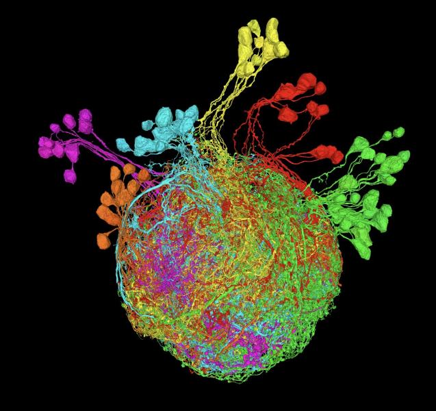
Premotor Neurons Controlling the Fruit Fly Leg
A reconstruction of premotor neurons controlling the fruit fly leg. Using an electron microscopy dataset of ultrathin sections of the Drosophila ventral nerve cord, researchers created a vivid display of the neural connections involved in fly leg movement. The structure of each neuron helps researchers determine their developmental lineages, represented by the different colors.
By Andrew Cook, Jasper Phelps, Anthony Azevedo, Ellen Lesser, Leila Elabbady, Brandon Pratt, Wei-Chung Allen Lee, John Tuthill, University of Washington and Harvard Medical School
Third Place
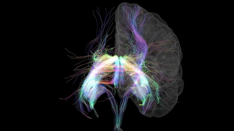
Memory Lanes
The hippocampus is the brain’s memory center. By combining two MRI scans, researchers can reveal the vast network of nerve fibers to and from the hippocampus—a wiring diagram for part of the brain. The axon fiber bundles are artificially colored depending on which direction they are heading. For a better sense of just how immensely complex the brain’s wiring is, this image represents less than 1% of the data collected.
By Tyler Ard, USC Stevens Neuroimaging and Informatics Institute
2022
Video Winners
Photo Winners
First Place
The Intersection of Memory and Memory
Two memories captured under the microscope. Peering into the hippocampus of a mouse using viral technology and optogenetics.
By Stephanie Grella, Boston University
Second Place
Mindmap – The Intricate Wiring of The Human Brain
Brain activity is orchestrated by propagating information between brain regions through fiber tracts, visualized via diffusion MRI tractography.
By Sahar Ahmad, Ye Wu, and Pew-Thian Yap, The University of North Carolina at Chapel Hill
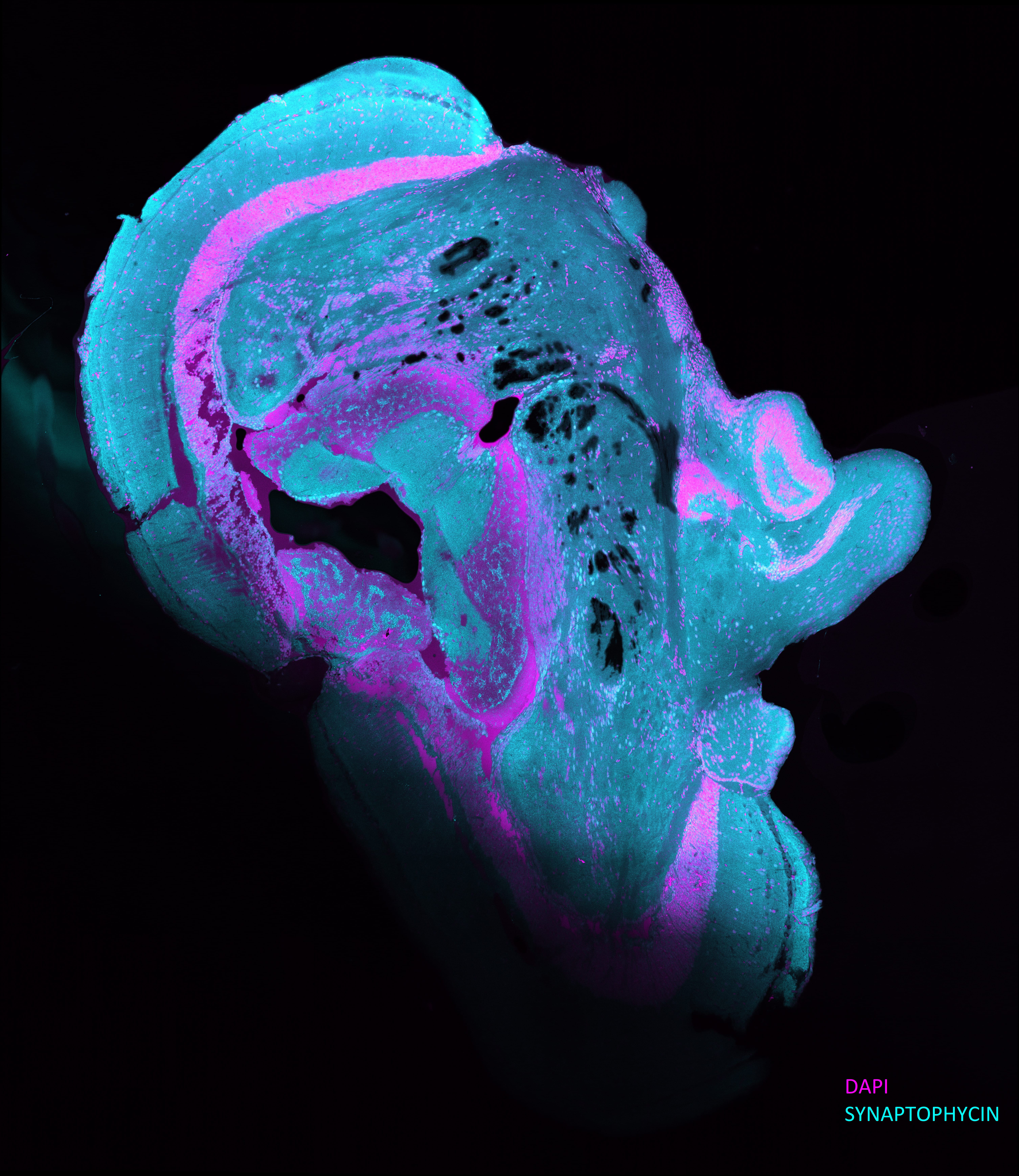
Third Place
Zebrafish Brain Thinking Abraham Lincoln
Image taken from a Zebrafish brain tissue section, synaptophysin as the primary antibody, Alexa555 and DAPI as secondary antibody, and looks like Abraham Lincoln's side profile.
By Esengül Öztürk, Çanakkale Onsekiz Mart University
2021
Download the 2022 BRAIN Initiative single page calendar(pdf, 5983 KB) featuring the top entries from the 2021 BRAIN Initiative Photo & Video Contest.
Video Winners
Photo Winners
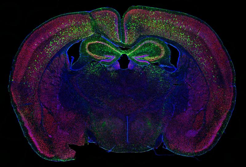
First Place
Thinking About a Greener Future
Mouse brain showing green AAV-transduced cells in the cortex and hippocampus. Neurons are labeled red and nuclei are blue.
By Allen Yen, Washington University School of Medicine
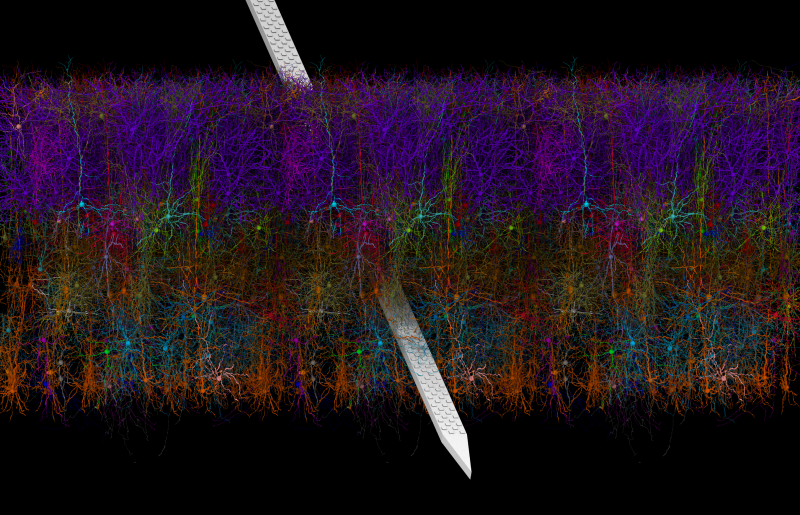
Second Place
Model of Mouse V1 with a Neuropixels Probe
This rendering of a model of mouse primary visual cortex with a Neuropixels probe was created using a new tool, VND (Visual Neuronal Dynamics).
By Barry Isralewitz, John Stone, Mariano Spivak, Kael Dai, Josh Siegle, Emad Tajkhorshid, and Anton Arkhipov, University of Illinois at Urbana-Champaign and Allen Institute
Third Place
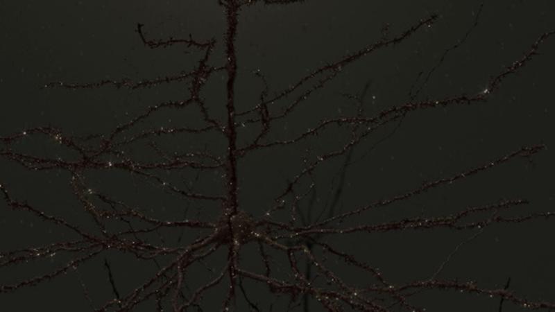
Neuron on Fire
Hippocampal CA1 pyramidal neuron in the mouse, recorded from the distal dendrite using patch clamp electrophysiology. The recorded location is visible as the gap in the dendrite. The neuron was filled with biocytin during recording and immunostained with streptavidin-647 post hoc. Confocal image was filtered using ImageJ.
By Olesia Bilash, New York University
2020
Download the 2021 BRAIN Initiative single page calendar(pdf, 2289 KB) featuring the top entries from the 2020 BRAIN Initiative Photo & Video Contest.
Video Winners
Photo Winners
First Place
Cortical Forest
Mouse Layer V cortical neurons eYFP-labeled (Thy1-H) and imaged after CLARITY processing of a whole brain. Maximum projection with depth color coding.
By Linus Manubens-Gil and Jim Swoger, Centre de Regulació Genòmica (CRG) and EMBL Mesoscopic Imaging Facility
Second Place
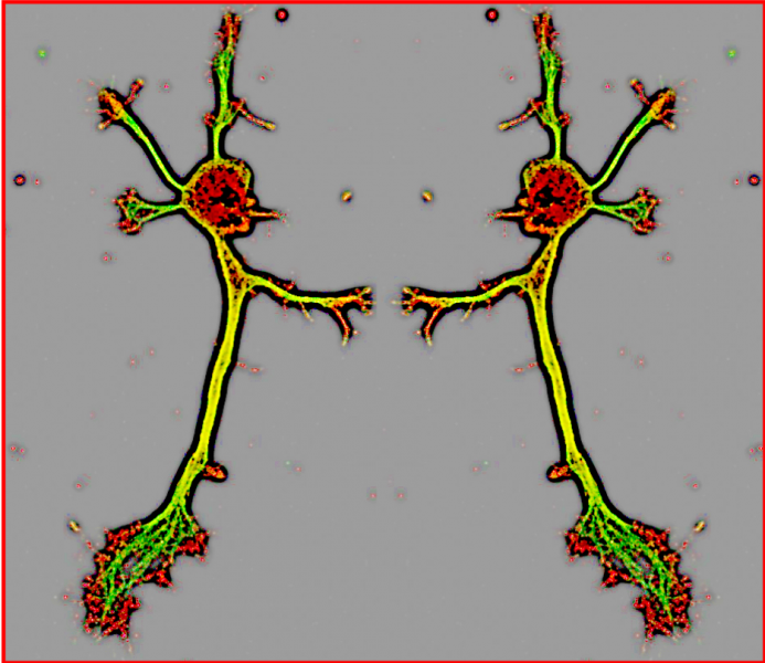
Radiating Neurons
4-week-old rat cortical neurons labeled for dendrites (red), axons (green), and nuclei (blue).
By Karthik Krishnamurthy, Davide Trotti, and Piera Pasinelli, Thomas Jefferson University
Third Place
The Ephemeral Hippocampus
The brain is everywhere to us neuroscientists. This exquisite 'hippocampus', with delicate dendrites, is actually a waterdrop captured at highspeed.
By Tallie Z. Baram, Jeremy Barry, and Joan Morris, University of California, Irvine, © 2017 Joan Morris
2019
Video Winners
Photo Winners
First Place
Light Me Up!
Light-based rendering of deep brain stimulation’s electrical excitation of neuronal fiber pathways to treat patients with traumatic brain injury.
By Andrew Janson, University of Utah Scientific Computing and Imaging Institute
Second Place
Dancing Devils
Mouse hippocampal neuron stained for f-actin (red) and tubulin (green).
By Sharada Tilve, NIH National Heart, Lung, and Blood Institute (NHLBI)
Third Place
Neural Circuit in The Storm
3D image of parvalbumin+ neurons (red, neurites; green, presynaptic puncta) swimming through the waves of GAD1+ (cyan) neurons.
By Young-Gyun Park, Massachusetts Institute of Technology (MIT)